Restoring brain IL-15 signalling unlocks innate lymphoid immunity against brain metastasis
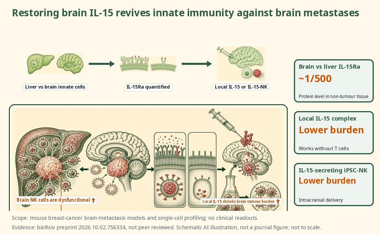
In a breast-cancer brain-metastasis model, group 1 innate lymphoid cells are profoundly dysfunctional and fail to control tumour, which the authors trace to severely restricted IL-15: IL-15Rα protein is about 500-fold lower in brain than liver; local IL-15 complex restored innate-cell accumulation, cytotoxicity and residency and cut metastatic burden independent of T cells, and intracranial IL-15-secreting iPSC-NK cells also reduced tumour burden.
Preprint. This study has not yet been peer reviewed; findings may change.
Using single-cell RNA sequencing and functional depletion, the authors compared group 1 innate lymphoid cells (NK cells and ILC1s) in breast-cancer brain metastasis (BCBM) with those in liver. In the liver, diverse, functionally competent group 1 ILC populations supported excellent metastatic control; in the brain the cells showed limited diversity and profound tissue-specific dysfunction, and their depletion had no effect on tumour burden despite modest parenchymal infiltration. The dysfunction was attributed to severely restricted IL-15 availability, with IL-15Rα protein detected at about 500-fold lower concentration in the brain than the liver. Local IL-15 complex administration restored group 1 ILC accumulation, cytotoxicity and tissue residency and significantly reduced brain metastatic burden independent of T cells. Intracranial delivery of IL-15-secreting induced pluripotent stem-cell-derived NK (iPSC-NK) cells, already in clinical testing for non-small-cell lung cancer, significantly reduced tumour burden. Mouse numbers per group and quantitative burden reductions are not stated in the available preprint abstract.
Background and open questions
Brain metastasis is a common and often fatal complication of breast cancer, and the brain has long been considered an immunologically distinctive site where systemic immunotherapies frequently underperform. Natural killer (NK) cells and type 1 innate lymphoid cells (ILC1s), collectively group 1 innate lymphoid cells, are rapid, antigen-independent killers that contribute to control of metastatic disease in several organs. Whether these innate effectors can control tumour in the brain, and if not, why, has remained poorly defined.
A recurring puzzle in the metastasis field is that the same cancer cells can be efficiently controlled in one organ yet grow unchecked in another. The liver and the brain offer a sharp contrast: the liver hosts abundant and varied innate lymphoid populations, while the brain parenchyma is comparatively sparse in these cells and is shaped by a specialised microenvironment. The open question the authors pose is whether group 1 ILCs are simply absent or outnumbered in brain metastasis, or whether they are present but functionally disabled by something local, and if so whether that local deficit can be corrected to reawaken innate anti-tumour immunity.
Study design
The authors combined single-cell RNA sequencing with functional depletion models to profile group 1 ILCs in breast-cancer brain metastasis (BCBM) and to compare them directly with the same cell classes in liver metastasis. Single-cell sequencing characterised the diversity and transcriptional state of the innate cells in each tissue, while antibody-mediated depletion tested whether the cells were actually required to restrain tumour growth at each site. Dual immunohistochemistry was used to quantify IL-15Rα protein in brain versus liver, probing the cytokine support available to innate cells in each environment.
On the therapeutic side, the team pursued two complementary strategies to restore IL-15 signalling where it was found lacking. First, they administered an IL-15 complex locally to test whether supplying the cytokine could rescue innate-cell numbers and function and reduce tumour burden. Second, to move toward a translatable modality, they delivered IL-15-secreting iPSC-derived NK cells intracranially; this platform is already in clinical testing for non-small-cell lung cancer. Experiments were designed to isolate the innate contribution, including a demonstration that the IL-15 complex benefit occurred independent of T cells. Per-group animal numbers and dosing details are in the full preprint but are not captured in the available abstract.
Key results
Innate lymphoid cells are competent in liver but disabled in brain
Single-cell sequencing revealed a stark tissue contrast. In the liver, diverse and functionally competent group 1 ILC populations were associated with excellent control of metastasis. In breast-cancer brain metastasis, the same cell classes showed limited diversity and profound, tissue-specific dysfunction. The functional consequence was demonstrated directly by depletion: removing group 1 ILCs in the brain had no effect on tumour burden, despite the cells showing modest infiltration into the parenchyma. In other words, the cells were present but not contributing to tumour control, a phenotype of disablement rather than mere absence. This distinction matters therapeutically: a disabled cell that is physically present can in principle be re-activated in place, whereas an absent one would first have to be recruited or supplied.
A roughly 500-fold IL-15 deficit in the brain
To explain the dysfunction, the authors turned to IL-15, a cytokine essential for NK- and ILC1-cell development, survival and effector function that is typically presented bound to IL-15Rα. Quantifying IL-15Rα protein by dual immunohistochemistry, they found it present at about 500-fold lower concentration in the brain than in the liver. This dramatic local deficit provides a mechanistic candidate for why innate lymphoid cells arriving in the brain fail to mount an effective response: they are deprived of the cytokine support that sustains their killing programme. IL-15 trans-presentation by IL-15Rα on neighbouring cells is a central way NK and ILC1 cells receive survival and activation signals, so a deficit of that presenting receptor would be expected to blunt both their persistence and their cytotoxic output.
Local IL-15 restores killing and cuts tumour, without T cells
Supplying IL-15 locally reversed the dysfunction. Administration of an IL-15 complex restored group 1 ILC accumulation, cytotoxicity and tissue residency, and significantly reduced brain metastatic burden. Importantly, this benefit occurred independent of T cells, isolating the effect to the innate compartment and arguing that reawakening innate killers is sufficient to improve control of brain metastasis.
A translatable cell-therapy route
Finally, the team tested a modality closer to the clinic. Intracranial delivery of IL-15-secreting iPSC-derived NK cells, a platform already in clinical testing for non-small-cell lung cancer, significantly reduced tumour burden in the model. This couples the mechanistic insight, that the brain starves innate cells of IL-15, with a delivery strategy that both supplies effector cells and brings their own IL-15 support, pointing toward a route that could in principle be translated.
Mechanistic interpretation
Demonstrated in the paper: Group 1 innate lymphoid cells in breast-cancer brain metastasis are dysfunctional rather than simply scarce, and their depletion does not change tumour growth. This dysfunction tracks with an approximately 500-fold lower level of IL-15Rα protein in the brain than the liver. Restoring IL-15 locally, either as a complex or via IL-15-secreting iPSC-NK cells, re-establishes innate-cell accumulation, cytotoxicity and residency and reduces metastatic burden, and for the IL-15 complex this occurs independent of T cells.
Author hypotheses: The authors argue that tissue-specific differences in cytokine availability shape the diversity and disease-controlling capacity of group 1 innate lymphoid cells. They propose that the brain's severe IL-15 deficit is a principal cause of innate immune failure against intracranial tumour, and position localized IL-15 signalling as a compelling strategy to unlock innate immunity against brain metastasis and potentially other intracranial malignancies.
Limitations and uncertainties
- This is a bioRxiv preprint and has not been peer reviewed; conclusions and any numeric values may change on revision.
- The data are from mouse models of breast-cancer brain metastasis and tissue sections; the IL-15 threshold and therapeutic window in human brain metastasis remain to be established.
- Per-group animal numbers, quantitative tumour-burden reductions, dosing and follow-up are not stated in the available abstract.
- Systemic IL-15 can carry toxicity, and the feasibility, safety and durability of repeated intracranial delivery in patients are unaddressed here; the referenced iPSC-NK clinical testing is in a different disease (non-small-cell lung cancer).
Clinical and industry implications
The study reframes a hard clinical problem: rather than the brain simply lacking innate immune cells, it appears to actively disable the ones that arrive by withholding IL-15. That shifts the therapeutic target from recruiting cells to restoring their local cytokine support. Two routes are suggested. A local IL-15 complex could in principle be used to reawaken endogenous innate effectors, while IL-15-secreting iPSC-NK cells offer an off-the-shelf cell product that supplies both the effectors and their cytokine support in one dose, delivered where it is needed. Because the referenced iPSC-NK platform is already in clinical testing for non-small-cell lung cancer, there is a plausible translational path, though brain metastasis would require its own trials. The immediate contribution is mechanistic and preclinical: establishing a measurable, roughly 500-fold IL-15 deficit as a driver of innate immune failure in brain metastasis, and showing that correcting it locally can restore innate anti-tumour activity. Human validation of the IL-15 threshold, delivery feasibility and safety remain the key open steps.
Authors and source
Timothy P. McMullen (University of California, Irvine) is first author. Co-authors include Pascal Naef, Erika Zagni, Lei Tian, Eva Zhao, Noah Wechter, Aaron J. Longworth, Isam Adam, Jacob Insua-Rodriguez, Hannah Savage, Isaac Hur, Sophia A. Garcia, Dennis Ma, Michael H. Liew, Sharmila Mallya, Kai Kessenbrock, Timothy E. O'Sullivan, Ahmed Mohyeldin and Jianhua Yu, at the University of California, Irvine. The corresponding author is Devon A. Lawson (University of California, Irvine). Declared funding includes the California Institute for Regenerative Medicine (CIRM), multiple National Institutes of Health grants, the George E. Hewitt Foundation for Medical Research, a Canadian Institutes of Health Research postdoctoral fellowship, the Mark Foundation for Cancer Research and others. The authors declared no competing interests.
Preprint (not peer reviewed): https://doi.org/10.64898/2026.10.02.756334 ; https://www.biorxiv.org/content/10.64898/2026.10.02.756334v1 . Posted 10 October 2026; the posted copyright notice reserves all rights.
Numbers are as reported by the authors. Corrections: contact@
Related science
In vivo BCMA CAR-T phase 1: 15 months of follow-up, one ongoing sCR
Green CAR-T cells attacking a red myeloma cell at right
Synthetic TCR library: on average only 56% of VDJdb TCRs reproduce their annotated reactivity
Green peptide-MHC complexes binding synthetic TCRs, with one non-reproducing case marked in red
Targeting ZMYND8 increases effector-like exhausted T cells in mice and enhances immunotherapy responses
Green immune cells attacking red tumour cells
One email, with links to every paper. Reports and custom landscapes: contact@inlightbio.com.
Opens your email app with a pre-filled request; we add you by hand. Free, unsubscribe any time.